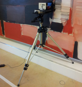
Raman spectroscopy is a vibrational spectrosccopy providing complementary information to FTIR. Generally it provides the identification of metal-oxide pigments (mainly transparent in the mid and near infrared range). The main limitation of Raman is related to the fluorescence emission (generated by the laser excitation) that may compete with the scattering phenomena covering all the vibrational signatures. To overcome this drawback, a multiple laser excitation is generally required thus favoring the longer wavelength lines for organic-containing matrices (varnished/unvarnished paintings, oil paints, manuscripts, etc..) and exploiting the higher energetic ones for inorganic-based substrates (ceramics, bronzes, etc, …)
The portable Raman spectrometer Rigaku Xantus-2, available at CNR-ISTM, has two different lasers operating at 785 and 1064 nm allowing the most suitable choice of laser excitation. For 785 nm laser, the power varies from 30 to 490 mW with a spectral resolution between 7 and 10 cm-1, and the detector is a CCD cooled by a Peltier system, while for the 1064 nm laser power varies from 30 to 490 mW, the spectral resolution between 15 and 18 cm-1 and an InGaAs detector is used. The spatial resolution is about 4 mm2.
The portable micro-Raman by Jasco is equipped with a Nd:YAG laser source emitting at 532 nm. The laser radiation is focused through an optical fibre into a JASCO RMP-100 microprobe equipped with an Olympus objective (50× or 20×). In the probe, a beam splitter focused the radiation co-axially into the objective, and the image of the irradiated sample is visualised by a CCD camera. The laser beam passed through a cut-off filter and a notch filter that eliminated the Rayleigh scattering of the excitation line. The backscattered Raman light is collected (at 180°) by a 2-m-long optical fibre with a diameter of 200 μm, and led to the Czerny–Turner polychromator (100 mm focal length). The detector is a 1024 × 128 pixel CCD ANDOR detector kept at −50° C with a Peltier cooler. The spatial resolution is 100 μm (20x objective) and the spectral resolution is about 10 cm-1.
References:
C. Miliani, F. Rosi, B.G Brunetti, A. Sgamellotti, “In situ Non-invasive Study of Artworks: the MOLAB Multi-technique Approach”, Accounts of Chemical Research 43, 2010, pp. 728-738.
F. Rosi, V. Manuali, T. Grygar, P. Bezdicka, B.G. Brunetti, A. Sgamellotti, L. Burgio, C. Seccaroni, C. Miliani, “Raman scattering features of lead pyroantimonate compounds: implication for the non-invasive identification of yellow pigments on ancient ceramics. Part II. In-situ characterization of Renaissance plates by portable micro-Raman and XRF”, Journal of Raman Spectroscopy, 42, 2011, pp. 407–414.
F. Rosi, C. Miliani, C. Clementi, K. Kahrim, F. Presciutti, M. Vagnini, V. Manuali, A. Daveri, L. Cartechini, B.G.Brunetti, A.Sgamellotti, “An integrated spectroscopic approach for the non invasive study of modern art materials and techniques” Applied Physics A Volume 100, (2010), pp. 613.
