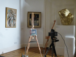
The performance of multispectral photographic imaging for this project aims at supplying compositional maps highlighting similitudes and/or differences in the pigment distribution on the paintings surface. This entails taking shots in visible light, UV Fluorescence, photographic IR, Reflected UV, using a photo camera Hasselblad 500 C, with a Leaf Aptus 65 digital camera back. The system is piloted by the LC11 program on Mac-Pro, and produces Raw images of 57.8 MB. Image calibration is guaranteed by including a set of 8 Spectralon Labsphere references in each shot, scaled from white to black.
The light source for the visible and infrared shots consists of two 800 W halogen lamps, while for the ultraviolet, both in fluorescence and reflected, a “Labino” Midlight/UV-A lamp peaked at 365 nm is used.
IR shots entail placing a Kodak 87 filter in front of the camera lens, after removal of the low-pass filter placed in contact with the photo camera’s sensor. A Schott KV450 filter is used for UV Fluorescence, while for Reflected UV a Kodak 18A filter is used, both maintaining the camera in the setup for visible shots, that is without removing the low-pass from the sensor.
The images in the various wavelengths, acquired without variation of position or enlargement, are calibrated in RAW format and then processed as Tiff. Combining in Photoshop the visible and near infrared photographs provides Infrared False color images, while the same procedure combining the visible with Reflected UV gives UV False Color images.
Another technical innovation, studied and applied for the first time during this project, is Infrared False Color Microphotography. A Sony DSC–F717 photo camera is mounted by means of an adaptor on the binocular optical microscope, taking advantage of the camera’s feature which permits the mechanical displacement of the low-pass filter, without changing the frame position and with autofocus. The re-assembly of the visible and infrared images in False Color is then done following the same procedure as described above. This technique, which permits observation and documentation with enlargements up to 20X, provides a particularly precise means of studying the morphology of details of the painted surface.
Comparison of all of these multispectral photographic images with specific reference standards may provide information on the different material distribution within the painted surface.